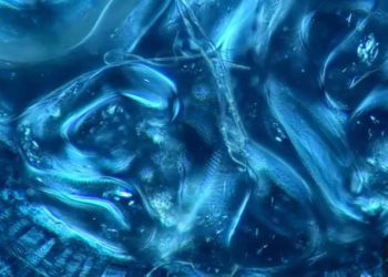
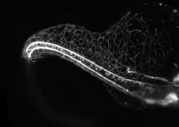
Each year Nikon offers up the very best of video microscopy in its Small World in Motion competition, where researchers submit their most stunning visual work. As a former bio-imaging researcher, the new approaches continue to amaze as the use of vectors to produce incredibly specific fluorescent proteins offers us new insight into cell and developmental biology. The progress is evident from comparing the sorts of work that won the contest in its first year (2011) to the current year’s winners below.
First place: Lateral line cells and melanocytes migrating in a zebrafish embryo
Second place: 12-hour time-lapse of cultured monkey cells labeled for plasma membrane (orange) and DNA (blue)
Third place: Sea anemone neurons and stinging cells showing their dynamic processes


